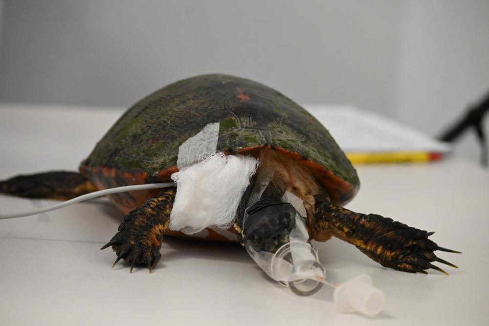
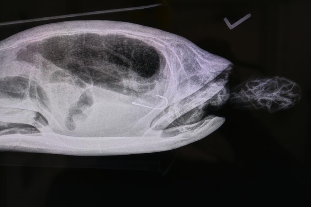
An American Red-bellied Turtle arrives at the hospital after being hit by a car. We’ve seen this before. In fact, we see it all too often. Their shell is fractured and requires surgical repair. On the surface, the solution seems simple; upon further diagnostic examination, our staff discovers the turtle also swallowed a fishing hook. This is a common theme in wildlife rehabilitation and conservation medicine- not everything is as it seems upon first glance.
Turtles’ and Tortoises’ shells are their bodies with intact nerve endings, blood vessels, and skeletal structures. Their shells are made of keratin- a tough, fibrous structural protein forming the primary building blocks for our hair and nails. The main concern with shell fractures is whether the injury caused spinal or vital organ damage. Turtles and tortoises are cold-blooded reptiles which means their metabolism is much slower than that of a bird or a mammal. As a result, shell fractures can take anywhere from 8 weeks to several months to properly heal. The recovery process becomes even more complex when underlying symptoms or issues are present. In this case, the underlying issue was a deeply embedded fishing hook in the esophagus. Again, upon first glance, it seemed like an easy extraction. As our veterinarians have done hundreds of times before, they made an incision to locate the hook and remove it; however, the location of the hook had advanced far beyond the accessible area. With little progress made, Dr. Tess McPheeters moved on to the shell fracture repair, knowing an efficient, successful hook removal would require more hands and additional equipment.
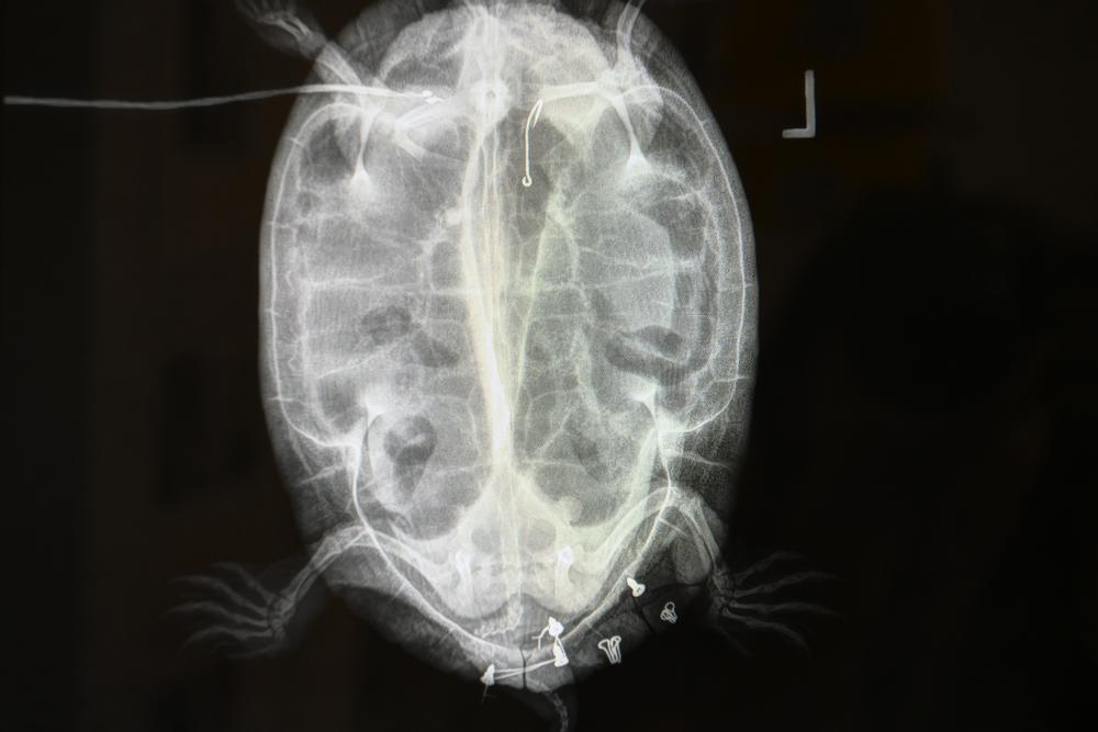
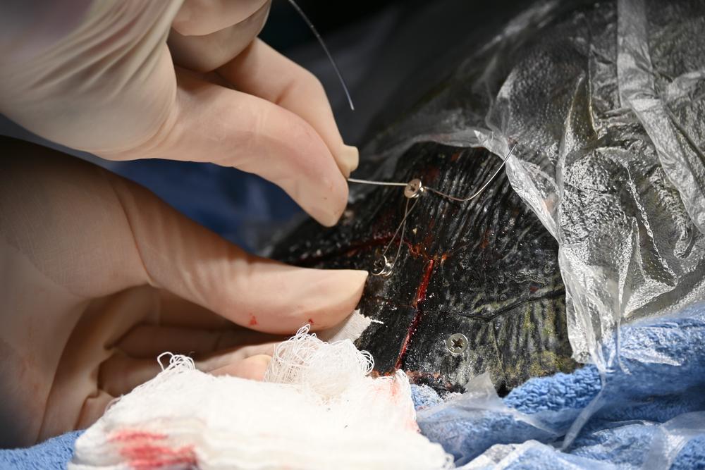
Thankfully, the shell fracture was small and located on the rear part of the carapace (upper shell) and did not impact the spine or any vital organs. Dr. Tess aligned the fractured scutes and shallowly advanced short screws to anchor the wiring that would gradually pull these pieces back together over time. Now, screws can hold the most tension and provide optimal healing results, but they aren’t the only option. In this case, Dr. Tess also utilized bra hooks secured through two-part epoxy application to anchor the shell where a screw might do more harm than good. Once all the anchors were in place, Dr. Tess weaved the wire between the screws and hooks. This wire will continue to be tightened, much like braces, during treatments and doctor checks throughout the healing process to allow the shell to keratinize over.
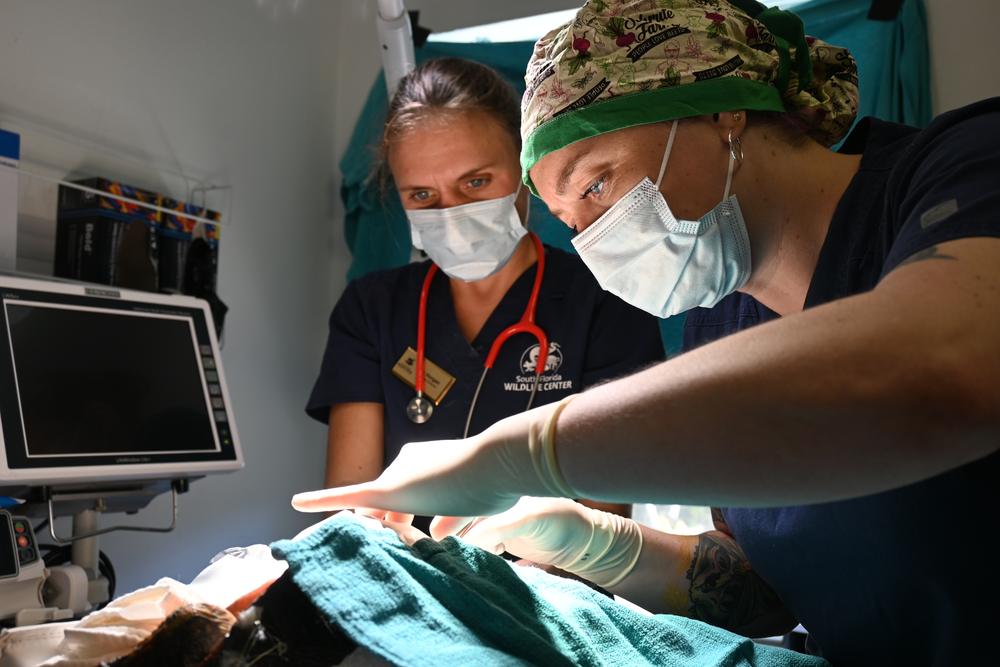
A few days later, after the turtle had stabilized and recovered from the shell fracture repair, our veterinarians, Dr. Molly Horgan, DACZM, and Dr. Tess McPheeters, attempted another hook extraction. This time they utilized our endoscopy machine to locate the exact location of the hook. It was positioned in the esophagus near the entrance of the stomach. After determining the precise location, they found the hook was significantly embedded in the esophagus and could not be simply grabbed and removed. They reopened the original incision at the base of the neck to retry the removal with better awareness of the exact location. Through careful maneuvering, Dr. Tess was eventually able to grasp the hook, freeing the barb from the internal esophageal lining, and back it out to the incision site. Dr. Horgan then clipped the barb from the end of the hook so it could be completely removed without causing additional damage. After the successful hook removal, Dr. Tess sutured the surgical site closed and placed the turtle in our Wildlife Ward to recover and begin the rehabilitation process. Our veterinarians expect the hardware to be removed after about 8 weeks if healing progresses appropriately. The American Red-bellied Turtle (26-5584) is currently healing well with a scabbed incision site and stable, immobilized shell fracture. Our staff will continue to administer supportive medication, nutrition, and therapies under close monitoring until the turtle is ready to graduate to an outdoor enclosure.
Wild animals can’t tell us what’s wrong. The members of the public who find them usually have limited, if any, history about what has happened to the animal. Unlike our cats and dogs, finders can rarely illuminate to our staff what wild animals have eaten, been exposed to, or how they’ve behaved over a period of time. This leaves room for a lot of unknowns and requires our staff to look at the entire picture through diagnostic testing, physical examinations, and trial and error to determine differentials and best treatment plans for each specific patient. With our patient intake load nearing 8,000 animals annually representing over 200 species, our staff have seen and experienced a multitude of situations requiring a plethora of approaches. Often, through discovering what doesn’t work, we’re able to implement what does to give these wild animals a chance at recovery to eventually return to their habitats where they belong.
If you find an animal you believe to be injured, sick, or orphaned, please call us for assistance at (954) 524-4302.
About South Florida Wildlife Center
Founded in 1969, South Florida Wildlife Center is one of the nation’s leading wildlife hospitals, providing rescue, rehabilitation, and release services for injured and orphaned native wildlife throughout South Florida. Caring for approximately 8,000 animals annually representing more than 300 species, the Center advances wildlife medicine, conservation, education, and community coexistence while training the next generation of wildlife professionals.
Love what you're reading?
Get the best of Weston delivered to your inbox every month — local stories, events, and good news worth sharing.
Subscribe for Free